This Breakthrough Method Turns Organs Transparent – Here’s How It Could Change Medicine Forever!


2025-08-17T22:46:58Z

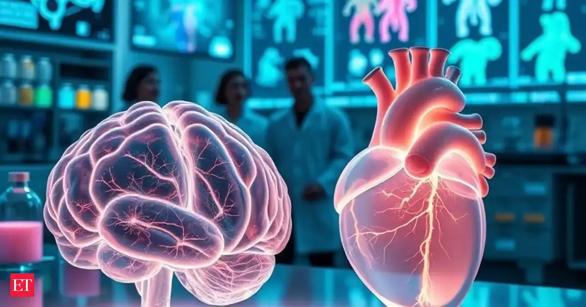
Imagine being able to see inside the human body without any incisions or damage—sounds like something out of a sci-fi movie, right? Well, this incredible innovation is no longer just a dream. Chinese researchers have developed a groundbreaking method that can make organs like the brain and heart transparent while preserving their original structure. This revolutionary approach could fundamentally change the landscape of medical imaging and disease research.
The method in question is called the ionic glass technique, and it's a game-changer for scientists who have long been stymied by the opaque nature of biological tissues. Traditionally, to get a detailed look at organs, scientists had to slice them into thin pieces, a process that destroyed the intricate connections within the tissues, making it nearly impossible to understand how everything works together in three-dimensional space.
With the ionic glass method, however, researchers can visualize entire organs in three dimensions without the need for dissection. This exciting advancement allows them to study crucial structures, such as neurons, blood vessels, and cardiac networks, without compromising their integrity.
Previously, biological tissues were a challenge to study due to their opacity, which blocked light and hindered the use of fluorescent dyes—crucial tools for highlighting cells and molecules. Existing clearing techniques often distorted samples, leading to expansion or shrinkage and the loss of delicate details. Even freezing methods, aimed at preserving samples, risked causing ice crystal damage, further complicating the research.
Now, thanks to a collaboration between Tsinghua University and several other institutions, including Beijing Chaoyang Hospital and Shanxi Medical University, researchers are using ionic liquids to create an “ionic glassy state” in organs. This innovative state allows light to penetrate easily, making it 2 to 30 times easier to see even faint biological signals. The team successfully examined the micro-connectivity of human neurons and even identified fine differences in impulse control compared to non-human brains.
The implications of this work are profound. By enhancing the visibility of rare proteins and neural pathways, this method could facilitate the creation of highly accurate 3D maps of entire organs—something that has eluded researchers for decades. According to the South China Morning Post, this advancement is touted as a potential game-changer, paving the way for breakthroughs in precision medicine and intelligent diagnostics.
Tsinghua University's social media page has even described their technique as providing “X-ray vision for tissues,” offering a built-in system to manage sample preparation, dye staining, and three-dimensional reconstruction. The future of medical research is looking brighter, and perhaps even clearer, thanks to this stunning innovation.
 James Whitmore
Source of the news: The Economic Times